
Amniotic Band Syndrome Presenting as Limb–Body Wall Complex:
A Rare Prenatal Diagnosis with Foetal Autopsy Correlation
Background: Amniotic Band Syndrome (ABS) is a rare congenital disruption sequence caused by rupture of the amniotic membrane, resulting in fibrous bands that entrap the developing fetus. Clinical manifestations range from isolated constriction rings to severe craniofacial defects, limb amputations and Limb–Body Wall Complex (LBWC). The reported prevalence varies from approximately 1 in 1,200 to 1 in 15,000 live births.
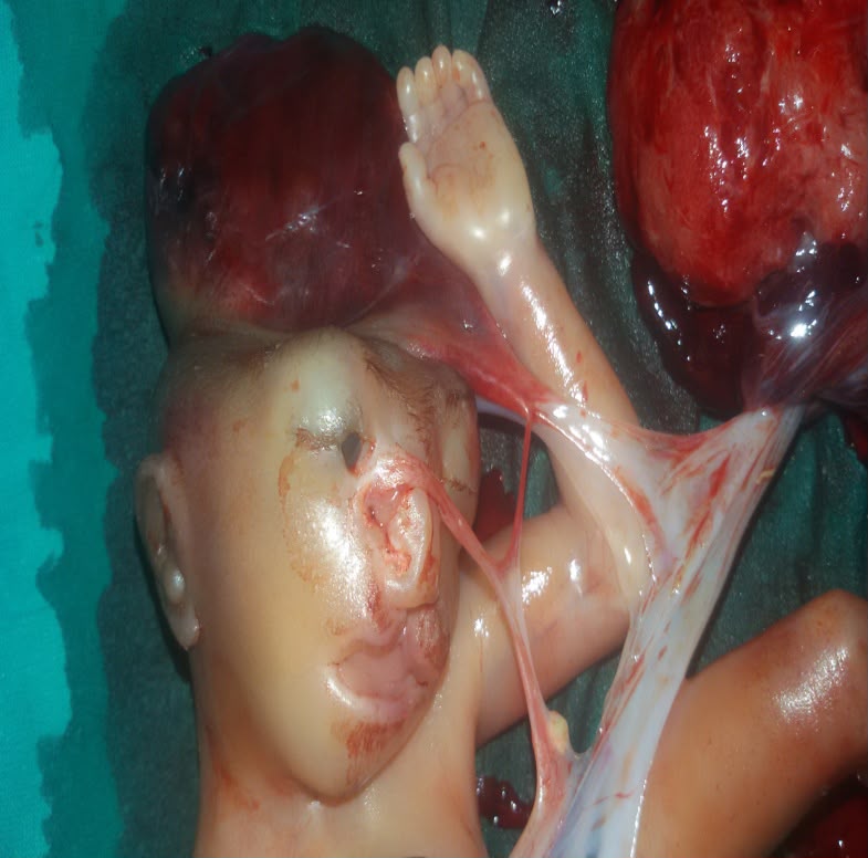
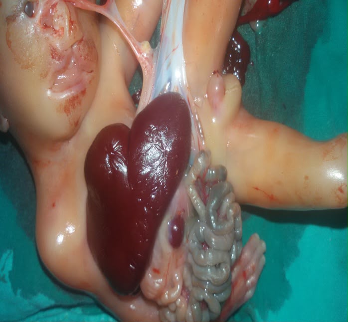
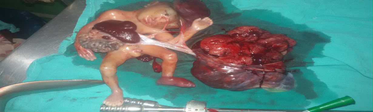
Case Presentation: A 23-year-old primigravida underwent routine anomaly scanning at 19 weeks of gestation, which revealed multiple severe congenital anomalies incompatible with life. Following detailed counselling and informed consent, pregnancy was medically terminated. Gross fetal examination demonstrated anencephaly, severe kyphoscoliosis, a large anterior abdominal wall defect with exteriorization of the liver, small intestine and urinary bladder, single umbilical artery, markedly short umbilical cord, cleft lip and multiple fibrous amniotic bands extending between the placenta and fetus. These findings confirmed Limb–Body Wall Complex associated with severe Amniotic Band Syndrome.
Conclusion: Early prenatal ultrasonography combined with fetal autopsy plays an essential role in diagnosis, parental counselling and recurrence risk assessment in pregnancies affected by severe congenital anomalies.
Keywords: Amniotic Band Syndrome, Limb–Body Wall Complex, Prenatal Diagnosis, Foetal Autopsy, Congenital Malformations.
Introduction
Amniotic Band Syndrome (ABS) is an uncommon congenital disruption sequence characterized by fibrous amniotic bands that entrap fetal structures, resulting in a wide spectrum of congenital anomalies. Clinical manifestations range from isolated limb constriction rings to severe craniofacial abnormalities and complex body-wall defects. The estimated prevalence varies from 1 in 1,200 to 1 in 15,000 live births.
Advances in prenatal ultrasonography have enabled early recognition of fetal anomalies, allowing timely counselling and appropriate obstetric management. Correlation between antenatal imaging and fetal autopsy remains invaluable for confirming the diagnosis and understanding the underlying pathogenesis.
We report a rare case of severe Amniotic Band Syndrome diagnosed at 19 weeks of gestation and confirmed by fetal examination following medical termination of pregnancy.
Case Report
A 23-year-old primigravida conceived spontaneously following a non-consanguineous marriage. She presented for a routine second-trimester anomaly scan at 19 weeks of gestation.
Ultrasonography demonstrated multiple severe congenital anomalies suggestive of a lethal fetal malformation. Following detailed counselling regarding fetal prognosis, pregnancy termination was performed after obtaining informed written consent.
Gross examination of the aborted male fetus revealed:
- Anencephaly
- Severe kyphoscoliotic short spine
- Extremely short neck
- Large anterior abdominal wall defect
- Exteriorized liver
- Exteriorized small intestine
- Exteriorized urinary bladder
- Gangrenous bowel loops
- Single umbilical artery
- Markedly short umbilical cord
- Wide cleft lip
- Multiple fibrous amniotic bands
- Swollen left foot
The external ears were normally developed. The palate, tongue and mandible appeared normal despite the presence of a wide cleft lip.
The fetus remained attached to the placenta through a markedly short umbilical cord and multiple fibrous amniotic bands. Thin translucent bands extended from the ruptured amnion to the fetal head, abdominal wall and exposed abdominal viscera.
Based on the prenatal ultrasound findings and gross pathological examination, a diagnosis of Limb–Body Wall Complex secondary to severe Amniotic Band Syndrome was established.
Discussion
The present case demonstrates one of the most severe phenotypic presentations of Amniotic Band Syndrome. The marked variability in clinical manifestations suggests that no single pathogenetic mechanism can explain the complete spectrum of abnormalities.
Amniotic Disruption Theory (Torpin)
Torpin proposed that rupture of the amniotic membrane results in fibrous bands arising from the chorionic surface. These bands entrap fetal structures and interfere with normal development. Early rupture produces severe craniofacial and visceral malformations, whereas later rupture usually causes isolated limb defects.
Vascular Disruption Theory (Van Allen)
This theory suggests that interruption of fetal blood supply during embryogenesis causes ischemia and tissue necrosis, resulting in congenital malformations.
Embryonic Dysplasia Theory
According to Streeter and McKenzie, localized defects in embryonic tissue development may independently produce constriction rings and associated anomalies without primary amniotic rupture.
Genetic Disorganization Theory
Donnai and Winter proposed that intrinsic genetic developmental abnormalities may explain rare ABS-like phenotypes presenting with body-wall defects, limb anomalies and unusual fetal malformations.
The present fetus demonstrated anencephaly, severe kyphoscoliosis, body-wall defect, exteriorized abdominal organs, markedly short umbilical cord and multiple fibrous amniotic bands. These findings strongly support the diagnosis of Limb–Body Wall Complex associated with early amniotic rupture.
Conclusion
Amniotic Band Syndrome should be considered whenever multiple asymmetric congenital malformations are identified on prenatal ultrasonography, particularly in association with body-wall defects and a short umbilical cord.
Early diagnosis enables accurate parental counselling, informed decision-making and appropriate pregnancy management.
Fetal autopsy remains indispensable for confirming prenatal findings and improving understanding of the complete spectrum of congenital abnormalities.
Current evidence suggests that the phenotypic manifestations of Amniotic Band Syndrome result from a combination of amniotic disruption, vascular disruption, embryonic dysplasia and genetic developmental mechanisms rather than any single pathogenic process.
Learning Points
- Amniotic Band Syndrome is a rare congenital disruption sequence.
- Limb–Body Wall Complex represents its most severe form.
- Early second-trimester anomaly scanning is crucial for diagnosis.
- Fetal autopsy significantly improves diagnostic accuracy.
- Multidisciplinary counselling is essential for recurrence risk assessment.
Patient Consent
Written informed consent for medical termination of pregnancy and publication of anonymized clinical details and clinical photographs was obtained from the patient's family.
Conflict of Interest
The author declares no conflict of interest.
Funding
No financial support was received for this work.
Acknowledgement
The author sincerely acknowledges the Department of Obstetrics & Gynaecology and the Department of Pathology for their support in the diagnosis and documentation of this rare congenital anomaly.